Human Osteology 3rd Edition Pdf – A classic in its field, human osteology has been used by students and professionals for twenty years. Now revised and updated in its third edition, the book continues to build the foundation of detailed photography and real-world scientific applications. New information, expanded coverage of existing chapters, and helpful additional images make this book invaluable for current work in the classroom and field.
Osteologists, archaeologists, anatomists, forensic scientists, and paleontologists will all receive accurate practical information on identifying, recovering, and analyzing and reporting human skeletal remains and making accurate deductions from those remains.
Human Osteology 3rd Edition Pdf

This field guide has been designed with practice as its main guide. Details of each bone 1) physiological features useful for identification, 2) other elements that can be confusing to the bone, 2) lateral instructions, 3) pictures of various stages of development, 4) useful information for age, and 5) time summary. Development. A concise description and subheading format helps to get information quickly.
Anatomy: Atlas Of Human Anatomy. Vols. 1 3. Vol. 1, Osteology, Arthrology And Syndesmology, Myology (317 Pp.); Vol. 2, Splanchnology, Ductless Glands, Heart (229 Pp.); And Vol. 3, Nervous System, A Ngiology, Sense
Principles of Bone Biology provides the most comprehensive reference for the study of bone biology and related diseases. This is an important resource for anyone involved in the study of bone biology. Bone research has received considerable attention in recent years, primarily because of the general public health impact of osteoporosis and related bone disorders. A classic in its field, human osteology has been used by students and professionals for twenty years. Now revised and updated for the third edition, the book continues … read more
A classic in its field, human osteology has been used by students and professionals for twenty years. Now revised and updated in its third edition, the book continues to build the foundation of detailed photography and real-world scientific applications. New information, expanded coverage of existing chapters, and helpful additional images make this book invaluable for current work in the classroom and field. Osteologists, archaeologists, anatomists, forensic scientists, and paleontologists will all seek practical information in the correct identification, restoration, and analysis and reporting of human skeletal remains and to make correct deductions from those remains.
Osteologist Team D. White, Michael T. Contains hundreds of amazing images in exquisite detail showing the maximum amount of anatomical information from the world-renowned and best-selling team of Black and photographer Peter A. Folkens. Updated case study examples showing life-size images of skeletal sections for easier learning and reference
Undergraduate and graduate students study human skeletal anatomy in physical anthropology, archeology, and medical school courses targeting the needs of coronary and forensic pathology; An essential basic reference and field guide for osteologists and critics, forensic scientists, paleontologists, and archaeologists.
Introduction To Human Osteology
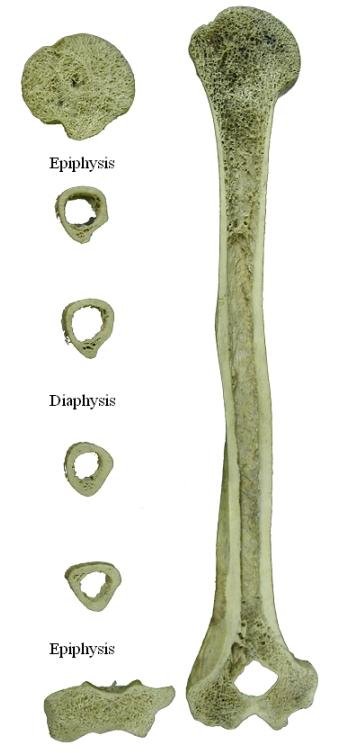
Introduction No. 3 Introduction No. 2 Introduction No. 1 Introduction 1.1. Human Osteology 1.2. Study guide 1.3. Teaching Osteology 1.4. Resources for Osteologists 1.5. Learn Osteology 1.6. Working with human bones Chapter 2. Body vocabulary 2.1. Field of reference 2.2. Directional requirements 2.3. Body movement 2.4. General characteristics of bones 2.5. Use of prefixes and suffixes 2.6. Physical space 2.7. Terms related to the form Chapter 3. Bone biology and differentiation 3.1. Changes 3.2. Some facts about bones 3.3. Bone is a component of the musculoskeletal system 3.4. Gross anatomy of bones 3.5. Molecular structure of bone 3.6. Bone Histology and Metabolism 3.7. Bone growth 3.8. Morphogenesis 3.9. Fixation of bones Chapter 4. Skull 4.1. Skull management 4.2. Components of the skull 4.3. Growth and Architecture, Sin and Sinus 4.4. Skull direction 4.5. Craniometric landmarks 4.6. Learning Cranial Skeleton Anatomy 4.7. Front (Figure 4.13-4.16) 4.8. Paretal (Figure 4.17-4.18) 4.9. Temporals (Figure 4.19–4.21) 4.10. Hearing ossicles (Figure 4.22) 4.11. Occipital (Figure 4.23-4.24) 4.12. Maxillae (Figure 4.25) 4.13. Palatines (Figure 4.26) 4.14. Vomer (Figure 4.27) 4.15. Inferior nasal concha (Figure 4.28) 4.16. Ethmoid (Figure 4.29) 4.17. Lachrymal (Figure 4.30) 4.18. Nose (Figure 4.31) 4.19. Zygomatics (Figure 4.32) 4.20. Sphenoid (Figures 4.33–4.36) 4.21. Mandible (Figure 4.37–4.39) 4.22. Measurement of the skull: Craniometrics 4.23. Gastrointestinal symptoms 4.24. Exam 5. Teeth 5.1. Dental form and function 5.2. Dental terminology 5.3. Teeth 5.4. Dental development 5.5. Identification of teeth 5.6. What kind of teeth are there? (Figure 5.5) 5.7. Are the teeth permanent or deciduous? (Figure 5.6) 5.8. Upper or lower teeth? 5.9 What is the position of the teeth? 5.10 teeth from the right or left? 5.11. Dental measurements: Odontometrics (Figure 5.21) 5.12. Nonmetric dental features Chapter 6. Hyoid and Vertebrae 6.1. Hyoid (Figure 6.1) 6.2. General characteristics of vertebrates 6.3. Cervical vertebrae (n = 7) (Figure 6.2 and 6.6) 6.4. Thoracic spine (n = 12) (Figures 6.3 and 6.8) 6.5. Hands (n = 5) (Figures 6.4, 6.10, and 6.11) 6.6. Measurement of the spine (Figure 6.12) 6.7. Symptoms of the spine Nonmetric 6.8. Characteristics of the spine Chapter 7. Thorax 7.1. Sternum (Figure 7.1-7.2) 7.2. Ribs (Figure 7.3–7.6) 7.3. Functional features of the Thoracic skeleton Chapter 8. Shoulder Girdle 8.1. Clavicle (Figures 8.1–8.5, 8.10) 8.2. Scapula (Figure 8.6–8.11) 8.3. Functionality of the shoulder belt Chapter 9. Hands 9.1. Humerus (Figure 9.1–9.8) 9.2. Radius (Figure 9.7, 9.9–9.15) 9.3. Ulna (Figure 9.7, 9.16–9.22) 9.4. Characteristics of elbow and wrist function Chapter 10. Hands 10.1. Carpels (Figures 10.4–10.11) 10.2. Metacarpals (Figure 10.12–10.18) 10.3. Hand phalanges (Figure 10.12-10.14, 10.19-10.21) 10.4. Characteristics of hand work Chapter 11. Pelvis 11.1. Sacrum (Figure 11.1–1.5) 11.2. Calculation (Figure 11.6) 11.3. Os Coxae (vav-11.12) 11.4. Pelvis (Figure 11.13–11.14) 11.5. Features of Pelvic Girdle Belt Chapter 12. Legs 12.1. Femur (Figure 12.1–12.8) 12.2. Patel (Figure 12.9–12.10) 12.3. Tibia (Figure 12.11–12.17) 12.4. Fibula (Figure 12.18–12.23) 12.5. Characteristics of knee and leg function Chapter 13. Leg 13.1. Tarsal 13.2. Metatarsal (Figure 13.18–13.22) 13.3. Foot Phalanges (Figures 13.18–13.20, 13.26–13.27) 13.4. Characteristics of foot work Chapter 14. Biological and biomechanical conditions 14.1. Anatomical conventions 14.2. Biomechanical convention 14.3. Translate Figure 14.4. Cranium and mandible 14.5. Clavicle 14.6. Humerus 14.7. Radius 14.8. Ulna 14.9. Os Coxae 14.10. Time 14.11. Tibia 14.12. Fibula Chapter 15. Field Procedures of the Remaining Skeleton 15.1. Find 15.2. Find 15.3. Mining and reclamation 15.4. Transportation Chapter 16. Laboratory Procedures and Reporting 16.1. Configuration 16.2. Stability 16.3. Preparation 16.4. Recovery 16.5. Alignment 16.6. Acquisition Metrics and Analysis 16.7. Photography 16.8. Radiography 16.9. Microscope 16.10. Molding and Casting 16.11. Computer 16.12. Report 16.13. Curation Chapter 17. Ethics in Osteology 17.1. Ethics and Law 17.2. Honoring the dead: appropriate personal behavior 17.3. Speaking for the Dead: Ethics in Forensic Osteology 17.4. Care of the dead: Considerations in the care of the remains 17.5. Management of the Dead: “Repatriation” and the American Burial Retention and Repatriation Act 17.6. Humanitarian ethics 17.7. Code of Ethics and Ethics Chapter 18. Assessment of age, sex, height, race, and identity of individuals 18.1. Determination of accuracy, precision, and reliability 18.2. From Known to Unknown: Using Standard Series 18.3. Estimated age 18.4. Gender Determination 18.5. Estimated height 18.6. Estimated ancestors 18.7. Identification of individuals Chapter 19. Osteoporosis and teeth 19.1. Description and diagnosis 19.2. Skeletal injuries 19.3. Congenital abnormalities 19.4. Circulation disorders 19.5. Joint diseases 19.6. Infectious diseases and related symptoms 19.7. Metabolic disease 19.8. Endocrine disorders 19.9. Anemia and disorders of blood cells 19.10. Skeletal dysplasia 19.11. Neoplastic conditions 19.12. Toothache 19.13. Muscle tension markers Chapter 20. Modification of the skeleton after death 20.1. Fracture 20.2. Bone improvement by physiological agents 20.3. Modification of bone by non-human biological agents 20.4. Bone modification by humans Chapter 21. Population biology of the skeleton 21.1. Nonmetric Alchemy 21.2. Assessment of biological distance 21.3. Food 21.4. Disease and Population Chapter 22. Molecular Osteology 22.1. Example 22.2. DNA 22.3. Amino acid 22.4. Isotope Chapter 23. Forensic case studies 23.1. A lost 23.2 in Cleveland. Research 23.3. Item 23.4. Determination 23.5. Summary Chapter 24. Forensic case studies 24.1. Child abuse and skeletons 24.2. Found a lost child 24.3. Analysis 24.4. Summary of Chapter 25. Archaeological case studies 25.1. Background 25.2. Geography Carson Sink 25.3. Disclosure and Recovery 25.4. Analysis 25.5. Branch 25.6. Osteoarthritis 25.7. Physical anus of the shaft 25.8. Physical stress 25.9. Restoration of food 25.10. Future Chapter 26. Archaeological case studies 26.1. Cannibalism and Archeology 26.2. Cottonwood Canyon Site 42SA12209 26.3. Find 26.4. Analysis 26.5. Chapter 27. What is the osteological composition? Atapuerca 27.2. Find 27.3. Recovery 27.4. Paleodemography 27.5. Paleopathology 27.6. Functional and phylogenetic evaluation 27.7. Continuation of the mystery Chapter 28 Case Study Forest Reserve 28.1. Background 28.2. Find fossils 28.3. Geography, Geology, and Geology Aramis 28.4. Explore “Ardi” 28.5. “Ardi” recovered 28.6. Restore “Ardi” 28.7. Document “Ardi” 28.8. Learning “Ardi” 28.9. Publishing “Ardi” Appendix 1. How to create images Appendix 2. A Decision Tree (“Key”) Approach to tooth Identification Appendix 3. Online Resources for Human Osteology Glossary Index Bibliography
The Human Evolutionary Research Center (HERC), and Department of Integrative Biology, University of California at Berkeley, CA, USA use cookies to customize content, customize ads and improve user experience. By using our website,
Netter atlas of human anatomy 3rd edition, human development a cultural approach 3rd edition, atlas of human anatomy 3rd edition, human osteology white pdf, advanced human nutrition 3rd edition, mathematics a human endeavor 3rd edition, an introduction to human factors engineering 3rd edition pdf free, human osteology 3rd edition, human rights politics and practice 3rd edition, human anatomy 3rd edition, biophysical foundations of human movement 3rd edition, mathematics a human endeavor 3rd edition pdf
